

SXR – Surgical Radiographic Guidance System
Choose options
A patented intra-surgical radiographic guidance device engineered to standardize periapical radiographic geometry and enhance accuracy during implant surgery.
PREORDER – 25% discount
Estimated shipping: March 1, 2026 (may ship earlier)
Quality & Compliance
• Manufactured in an ISO 13485:2016–certified medical device facility
• FDA Class I (Exempt) intraoral radiographic instrument — 21 CFR §872.4565
• U.S. Patent Pending
• Proprietary radiographic geometry for accurate, reproducible intra-surgical verification
• Validated processes for manufacturing, sterilization and final inspection
What Is SXR?
Sniper Xray (SXR) is a compact, precision-engineered radiographic guidance system designed to provide stable, reproducible and distortion-free periapical imaging during implant placement.
The system consists of two coordinated components:
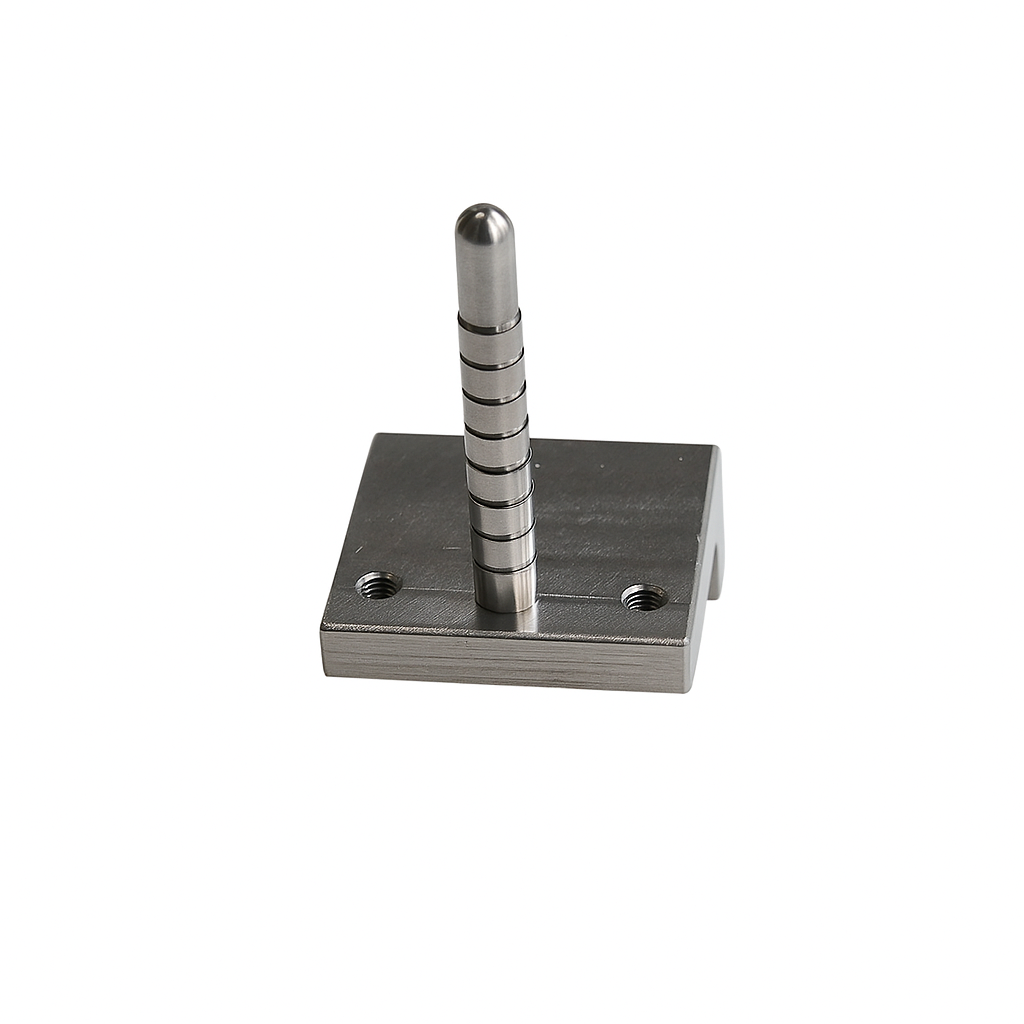
1. SXR Base
A rigid, fully autoclavable stainless-steel platform featuring dual locking bolts compatible with any XCP RINN bite block.
Key Features
• Three threaded positions to adapt probe angulation based on anatomy and film size
• Maintains true parallelism between probe and film
• Aligns the X-ray beam perpendicularly for distortion-free measurement
• Provides geometric stability essential for accurate intra-operative verification
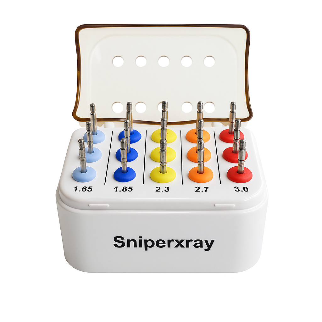
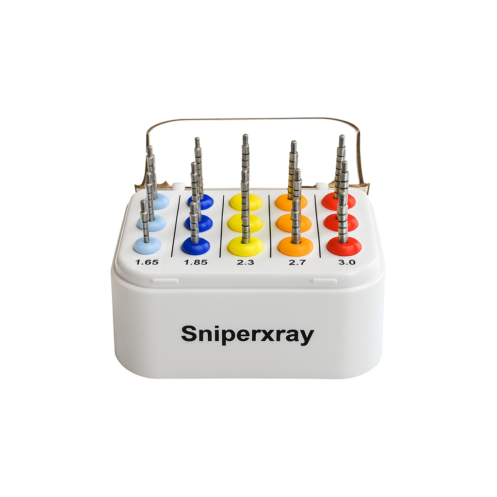
2. Depth Probes
A complete set of 15 stainless-steel depth probes, designed for intra-surgical radiographic depth assessment.
Diameters: 1.65, 1.85, 2.3, 2.7, 3.0 mm
Lengths: Short, Medium, Long
Radiopaque Markings
Short: 6, 8, 10, 12, 14 mm
Medium: 16, 18 mm
Long: 20, 22 mm
These markings provide precise intra-operative reference points for osteotomy depth verification and improved clinical decision-making.
How SXR Works
Select the probe
Choose diameter and length based on the final drill used in the osteotomy.
Assemble the system
Insert the SXR Base into an XCP RINN bite block and select the optimal threaded position.
Position intra-surgically
Place the probe gently into the osteotomy.
Stabilize
Have the patient bite lightly on gauze to immobilize the system.
Acquire the radiograph (PAR)
Probe and film remain parallel while the X-ray beam is perpendicular, producing standardized, reproducible geometry for accurate linear measurement.
Important:
Accuracy is operator-dependent. Optimal results require correct probe diameter, thread position and film placement. A brief learning curve is expected.
What’s Included
• 15 stainless-steel depth probes (1.65–3.0 mm)
• Short, Medium and Long lengths
• SXR Base with 3 threaded angulation positions
• Autoclavable storage box
• Radiopaque depth markings (6–22 mm)
• Printed Instructions for Use (IFU)
• Digital IFU available online
• Email-based clinical support
Preorder – Limited Release
SXR is available as a limited preorder with an exclusive 25% discount.
Estimated shipping: March 1, 2026 (may ship earlier).
You will receive email confirmation when your unit is ready for dispatch.


