University Testing & Proven Precision
Preliminary testing conducted by oral and maxillofacial radiologists at the University of Texas – San Antonio demonstrated Sniper Xray’s ability to maintain consistent projection geometry and deliver high precision in intraoperative periapical imaging.
From planning to real-time verification

The foundation of radiographic precision in implant dentistry
UNDERSTANDING PAR
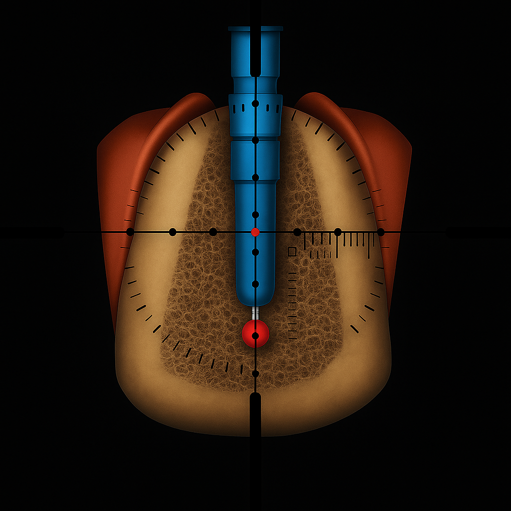
Periapical Radiography — What do we know?
Why PAR doesn’t work intra-operatively
Bridging preoperative planning and intraoperative verification
UNDERSTANDING SXR
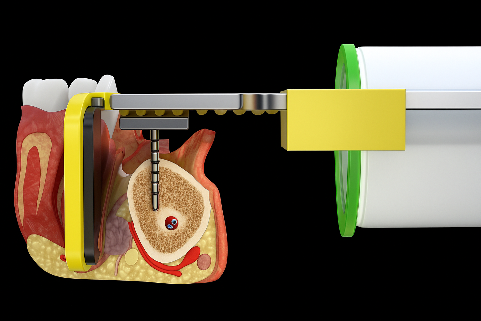
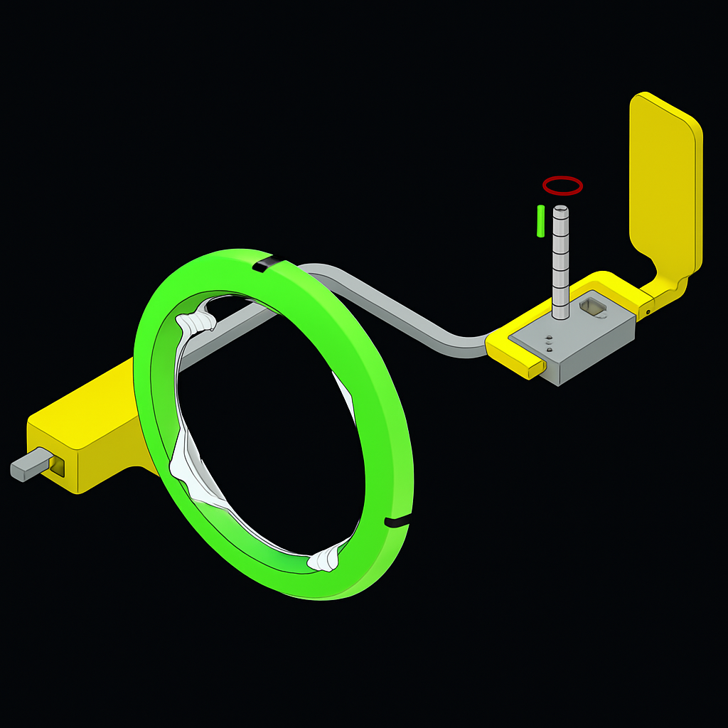
What is SXR?
How to Use SXR
What SXR adds
Advancing radiographic technology for safer, smarter surgery
CLINICAL INNOVATION
Computer-Assisted Verification
SXR Advantages
PRACTICAL VALIDATION OF SXR IN REAL CLINICAL SCENARIOS — THE SAME SYSTEM INCLUDED IN THE SXR EVALUATION UNIT
Clinical Cases
Case Reports
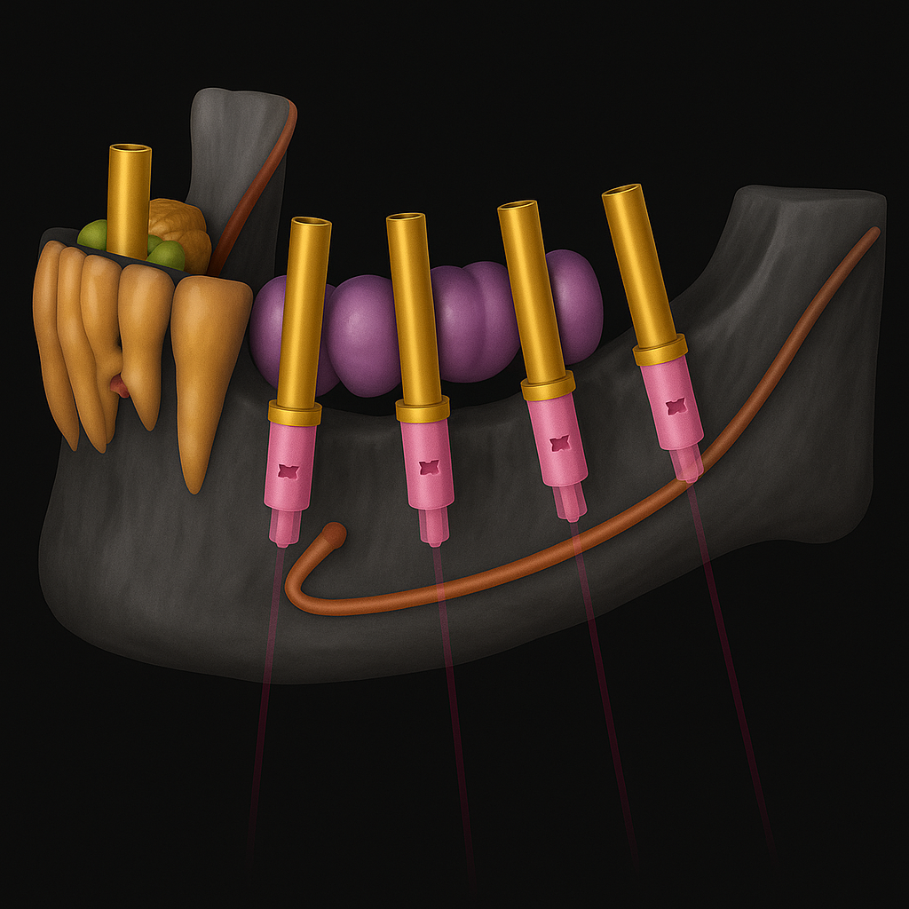
Sequential PAR Assessments to Enable Accurate Placement of Long Implants
Sequential PAR Assessments to Preserve the Inferior Alveolar Nerve

Developed by an oral surgeon with more than thirty years of experience in implant placement, SXR was conceived after a clinical event that revealed the need for safer, more accurate intra-surgical guidance.
Born from Experience, Built for Precision
Developed by an oral surgeon with more than thirty years of experience in implant placement, SXR was born from a difficult moment: an iatrogenic injury to the inferior alveolar nerve that created an unpleasant experience for both the patient and the surgeon.
That incident became the spark that led me to search for a better way.
After years of facing the same challenges every surgeon knows — intraoperative doubts, fear of perforation, lack of surface contact, and limited bone availability — I began to envision a tool capable of verifying, in real time, the depth, angulation, and relationship to nearby anatomical structures.
What began as a simple idea evolved through countless prototypes, trials, and refinements until the solution finally took shape.
That’s how SXR came to life: from experience, from necessity, and from the desire to make every procedure safer and more predictable.